
A Brave New World: Applying AI to Melanoma
“AI will change everything in the world—and melanoma is no exception,” says Dr. Allan C. Halpern, Chief of Dermatology at Memorial Sloan Kettering Cancer Center.
Halpern is a board-certified internist and dermatologist with a specialization in skin cancer, especially melanoma. Much of his clinical career has focused on early detection and management of melanoma, and he has pushed the field to adopt and leverage technology to better image and track peoples’ skin over time.
Halpern pioneered the use of a whole-body, 3-D imaging system to assist in the detection of changing moles and other skin lesions. The computerized system creates a baseline digital photographic record of the patient’s moles, searchable by size, color, and other factors; which can then assist the dermatologist in monitoring and assessing subsequent changes over time. This includes subtle changes that can be more difficult for dermatologists to catch but which are critical for identifying melanoma at the earliest stage possible. “This can help you identify the outliers,” explains Halpern. “In clinical practice, this technology isn’t being widely used yet, but we expect that this is where it’s going.” This work was first supported by an MRA-Industry Partnership Award, and has since been successfully commercialized.
According to the U.S. Preventive Services Task Force, limitations in clinician visual skin examinations can lead to both over treatment and over-diagnosis. For patients, this can mean unnecessary procedures, scarring, and even functional limitations.
One challenge to widespread adoption of this technology is that the field of dermatology doesn’t have standard image requirements. In response, Halpern, and others like him, are working hard to develop digital communication standards.
To help make this happen, Halpern leads the International Skin Imaging Collaboration (ISIC)—an academic and industry partnership designed to facilitate the clinical application of digital skin imaging in melanoma. The ISIC community invites people to download images and use them for educational purposes. They also host competitions to create algorithms to better spot early melanomas. “What’s exciting,” says Halpern, “is when we compared the algorithms against 510 clinicians including dermatologists from around the world, the algorithms outperformed the majority of them. While this ‘artificial experiment’ was performed without the clinicians interacting with the patients, it does demonstrate that current computer algorithms are already pretty powerful. As we give computers lots more data, including both images and clinical information, we anticipate that their performance will continue to improve.”
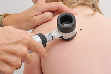
Dermascopes are helpful tools that allow clinicians to more closely inspect lesions on skin

Dr. Allan C. Halpern at 2019 MRA Scientific Retreat
While the vast majority of past MRA-funded research projects have focused on later-stage melanoma treatments, work like Halpern’s is laying an important foundation for future awards needed to move the field forward towards earlier detection and treatment. In particular, although early-stage melanoma is challenging for dermatologists to identify, AI might change that. Doing so would have significant benefits for patients as it would improve early diagnosis of melanoma.
“AI isn’t new,” Halpern says. “There have been multiple waves of AI. What’s different and exciting about this current wave is that it teaches computers like we teach kids, learning by example. This has been made possible because computers are much more powerful than they were years ago and now have nearly limitless storage.” Current AI uses what is known as “deep learning” one example of which are “convolutional neural networks.” These algorithms use multiple layers of analysis to learn directly from raw data. Such networks are particularly well suited for analysis of visual information, provided there are large enough collections of data for training. The recent availability of very large general image datasets give the algorithms a head start for analyzing new types of images, such as skin lesions.
Properly training AI for melanoma, however, will require a lot more imaging in clinical practice, collecting high-quality images, controlling for bias, and doing a better job applying what is learned so as to avoid unnecessary biopsies.
“It’s not a question of if we’ll use this technology, but when,” says Halpern. He is, however, quick to preface that this is not man against machine. AI, and those in support of it, are not trying to put people out of work. “What we want are better diagnoses,” says Halpern. “Even dermatologists do better when they know what the computer analysis is saying.”
Work like Halpern’s raises the bar of what is possible and begins to fill a critical niche: catching melanoma early when it is most treatable and survival rates are greatest. The implications are truly profound for patients and for professionals alike. “There are lots of opportunities for MRA to be involved in this work and to help implement it the right way,” says Halpern. Indeed, AI—much like MRA’s mission—requires collaboration. The groundwork has started; the revolution is here; the technology is capable; and the time is now.
2 U.S. Preventive Services Task Force. Screening for Skin Cancer: US Preventive Services Task Force Recommendations Statement. JAMA. 2016; 316(4): 429-435.
3 For more information, visit: www.isic-archive.com/#!/topWithHeader/wideContentTop/main
4 Halpern AC. Predicting the Future: AI, Machine Learning and Dermatology” at the 2019 American Academy of Dermatology Annual Meeting.